
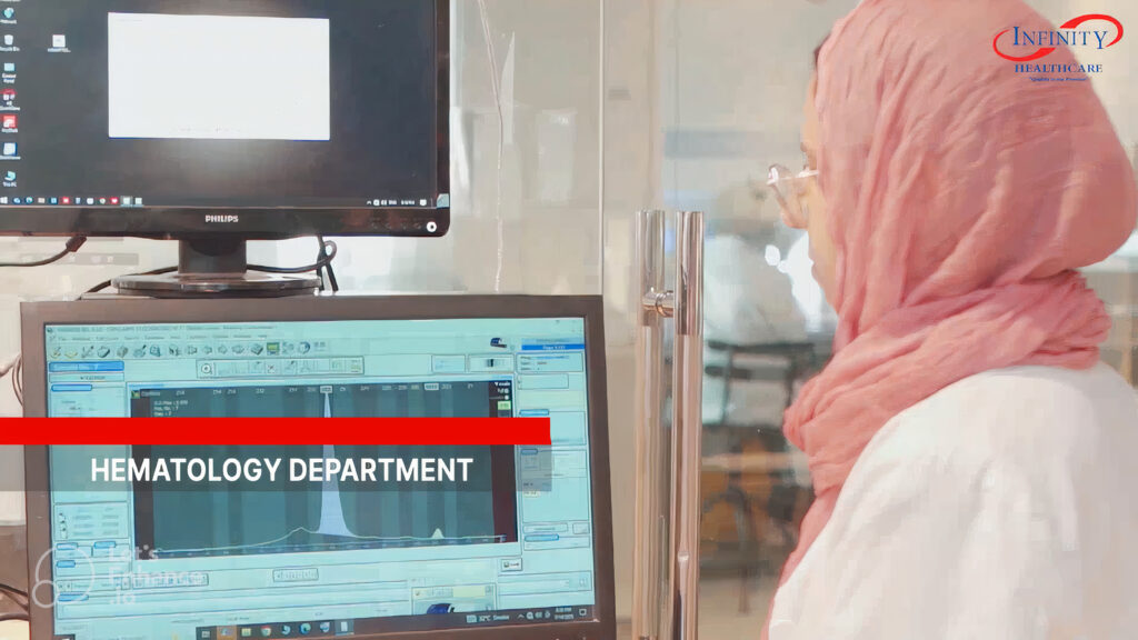
Hematology
Overview of Laboratory Hematology
Laboratory hematology encompasses all blood tests, and provides both quantitative and qualitative characteristics of analysed blood. The work of hematologists is usually divided into three areas: diagnosis of hematological disorders, management of hematological disorders, and blood transfusion. The diagnosis of hematological disorders is based on clinical history and examination, measurement of parameters in the blood, and microscopic examination of blood films, bone marrow aspirates and trephine samples. Automated machines measure multiple parameters in samples of blood; the most common are: Hb concentration, Red cell count, Hematocrit, red cell indices (MCV, MCHC), white cell count (WCC, DIFF), platelet count, and coagulation screening.
Blood Smear Examination
Examination of a blood smear provides information about the number, shape, and types of blood cells as part of a hemogram
It helps quantify leukocytes (DIFF), estimate platelet count, and detect morphological abnormalities linked to diseases. Blood films reveal changes in red blood cell shape and size (e.g., anisocytosis, poikilocytosis, macrocytosis) and abnormal white cells such as blasts in leukemia. Features like rouleaux formation may indicate abnormal plasma components, such as excess antibodies. Flow cytometry, used for immunophenotyping, is essential for diagnosing and monitoring hematological neoplasms by identifying and counting cells labeled with antibodies for specific antigens that indicate cell type, function, and maturity.
Bone Marrow Examination
Bone marrow samples are obtained by aspiration, biopsy, or both. A Giemsa-stained smear of aspirated cells helps identify marrow components, assess cellularity, detect fibrosis or neoplasms, and estimate iron stores. This is vital for diagnosing leukemia and monitoring treatment response. Trephine biopsies preserve tissue architecture, allowing evaluation of cellularity, reticulin, and cell distribution—especially important in fibrotic conditions like myelofibrosis or metastatic cancer, where aspiration yields few cells.